Eye Disease Diagnosis and Treatment
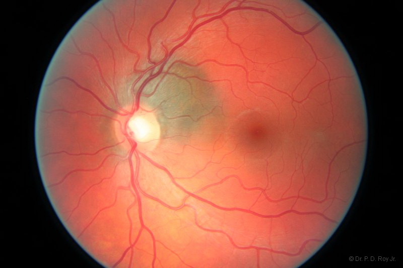
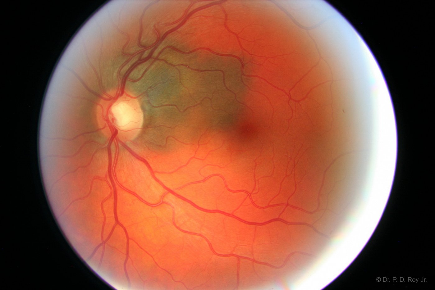
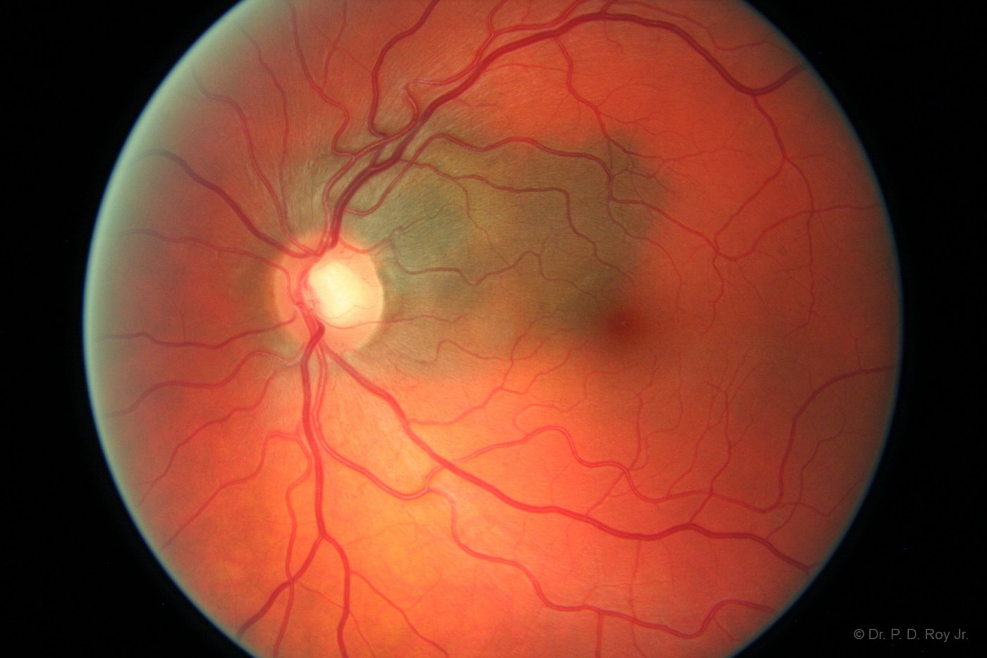
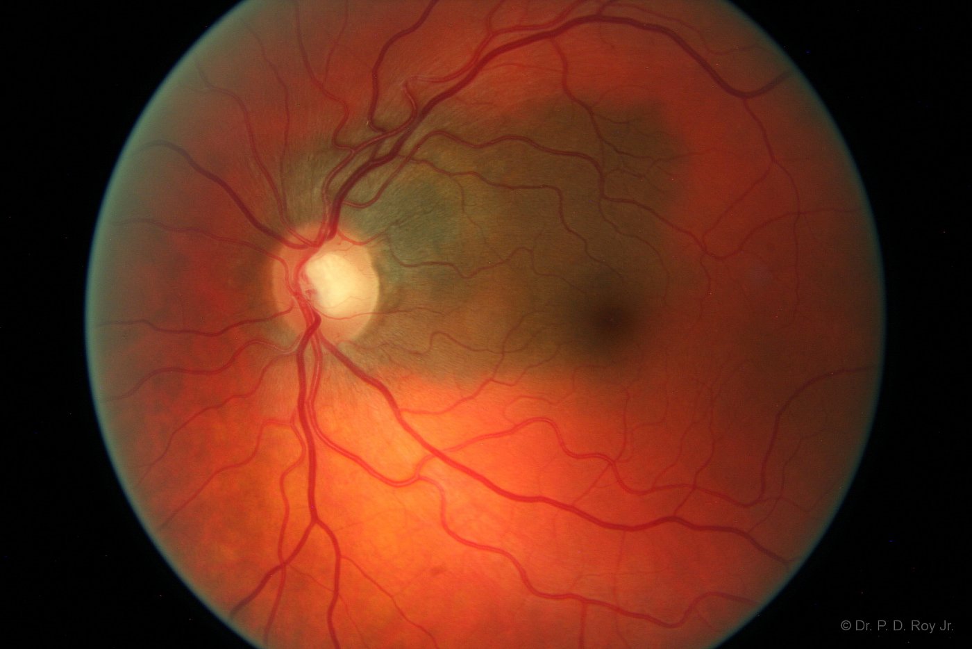
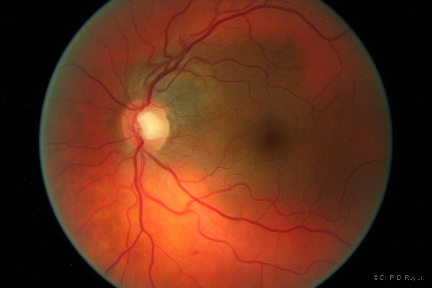
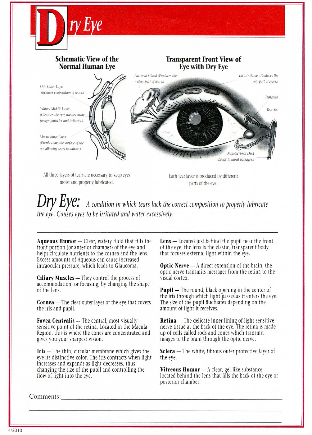
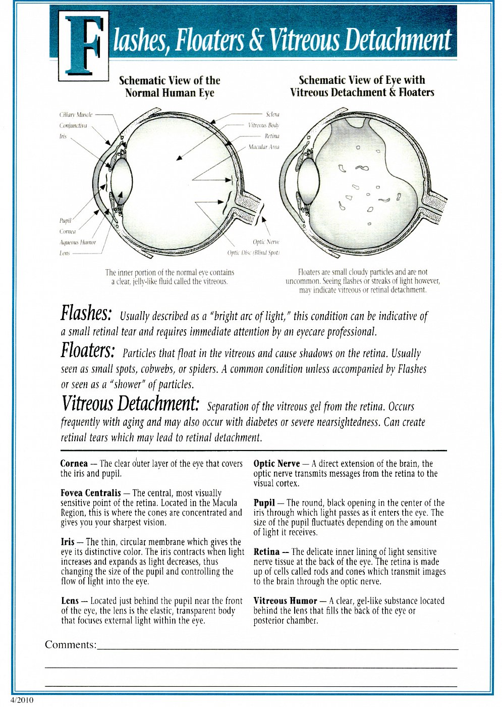
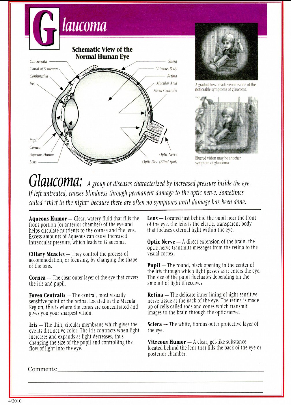
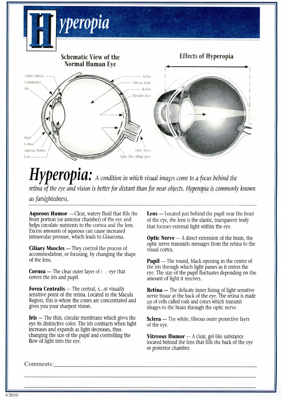
In every comprehensive eye exam, Dr. Roy will provide a thorough evaluation of your eye health. This allows for the detection of eye diseases such as cataracts, glaucoma, macular degeneration, retinal detachment, dry eye, etc. If you have a systemic condition such as diabetes, high blood pressure, or thyroid disease, you will be evaluated for any related eye disease. As a primary eye care provider, Dr. Roy is licensed to prescribe many ocular and oral medications. Should your condition(s) require surgical intervention or other care you will be properly co-managed with a sub-specialist in the area of your specific needs. The below photographs were made by Dr. Roy and show many common eye diseases.
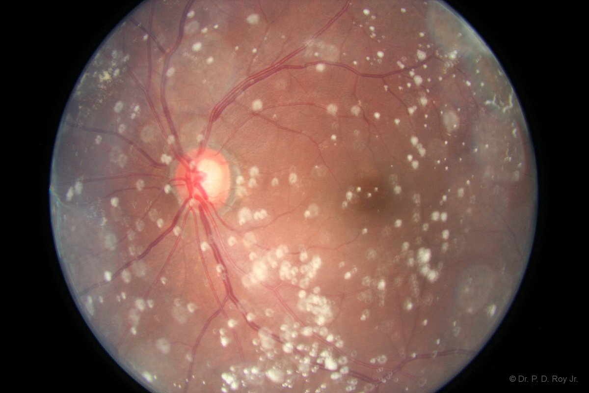
Asteroid Hyalites
Cataract
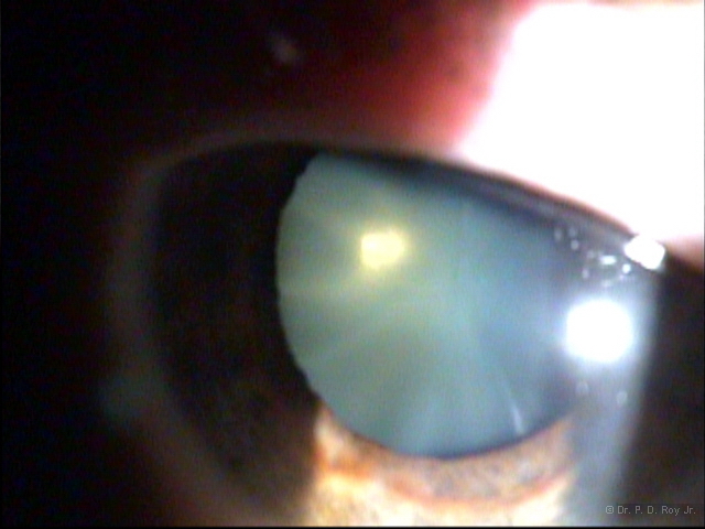
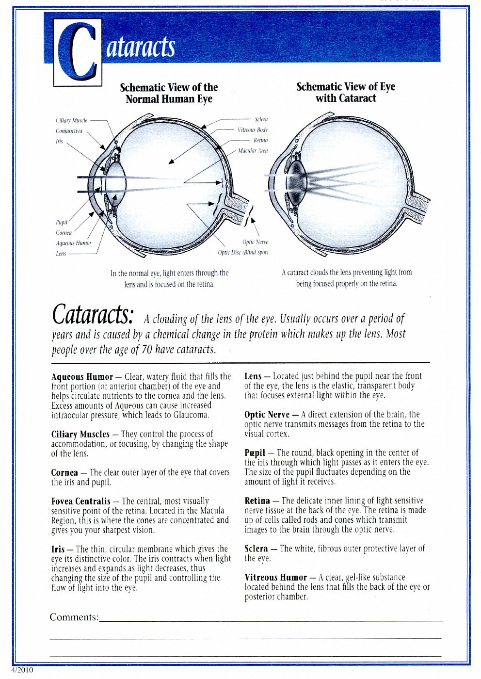
A cataract is a cloudy or opaque area in the normally clear lens of the eye. Depending on its size and location, it can interfere with normal vision. Most cataracts develop in people over age 55, but they occasionally occur in infants and young children. Usually, cataracts develop in both eyes, but one may be worse than the other. The lens is located inside the eye behind the pupil and iris, the latter of which is the colored part of the eye. Normally, the lens focuses light on the retina, which sends the image through the optic nerve to the brain. However, if the lens is clouded by a cataract, light is scattered so the lens can no longer focus it properly, causing vision problems such as blur. The lens is made of mostly proteins and water. Clouding of the lens occurs due to changes in the proteins and lens fibers. Cataracts generally form very slowly. Signs and symptoms of a cataract may include:
- Blurred or hazy vision
- Reduced intensity of colors
- Increased sensitivity to glare from lights, particularly when driving at night
- Increased difficulty seeing at night
- Change in the eye's refractive error, necessitating glasses prescription changes before the need for surgery.
Cataract
Before Removal
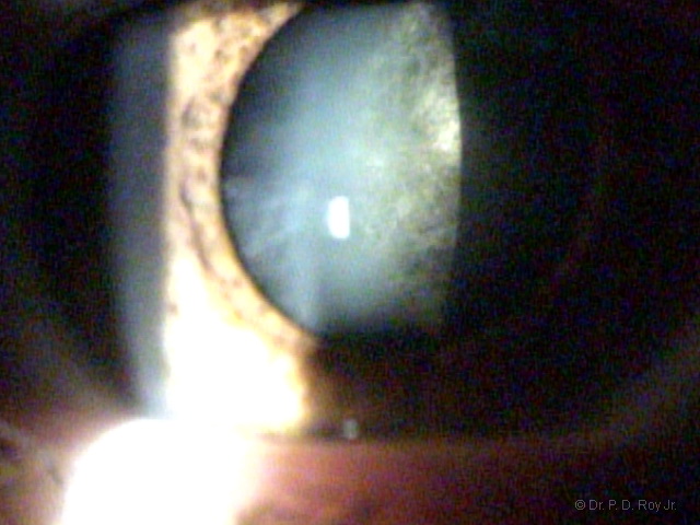
Cataract
Cataract Removed Intra-ocular Lens (IOL) Inserted (see edge of IOL). The IOL is clear unlike the cataract in above photograph.
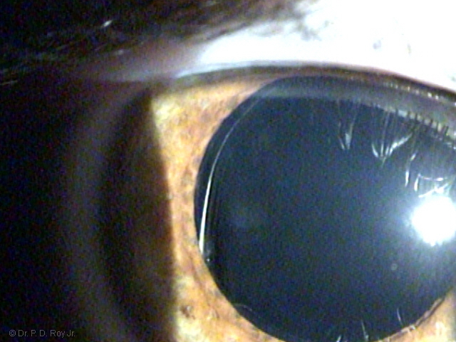
Photography by Dr. P. D. Roy, Jr. © All Rights Reserved.
Cataract
Intraocular Lens Behind Undilated Pupil
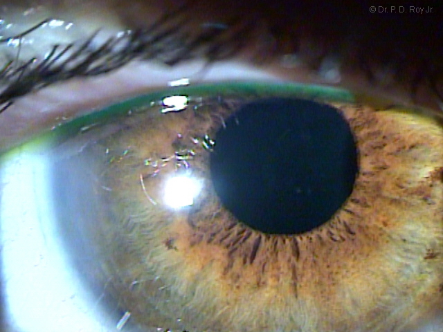
Old type of replacement lens
After Cataract is removed.
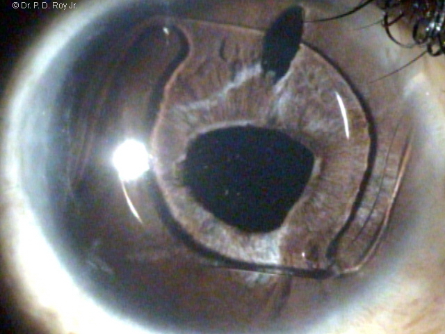
Corneal Abrasion
A corneal abrasion is a painful scrape or scratch of the surface of the clear part of the eye.
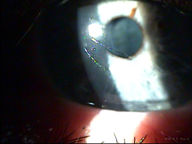
Corneal Transplant
This eye's pupil was obscured by the mostly opaque and cloudy cornea until the donor tissue was transplanted restoring the vision to the eye.
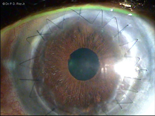
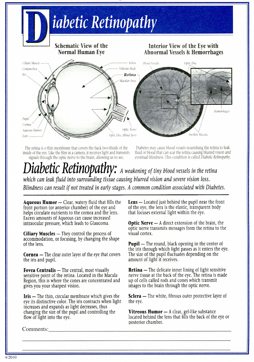
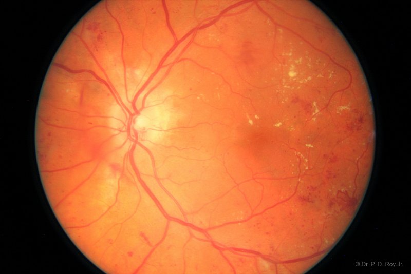
Diabetic Retinopathy
Seen here as the red and yellowish spots.
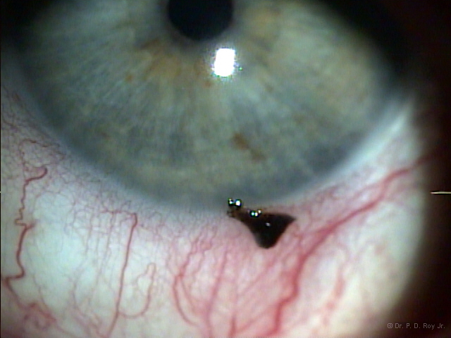
Foreign Body
Wearing eye protection (safety glasses with side shields) can often prevent scheduled office visits and safe costs and eyesight.
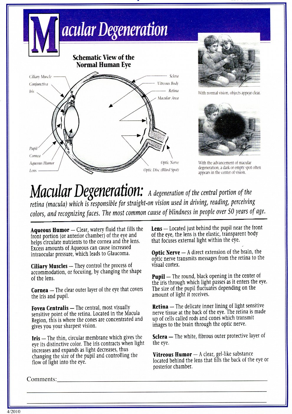
Macular Degeneration (dry)
Vitamins/antioxidants the AREDS 2 version has been shown to be helpful in reducing the progression of macula degeneration.
.jpg)
Macular Degeneration (wet)
This type of macula degeneration may be slowed in its progression with the placement of a special drug (commonly used since about 2005) into the eye.
.jpg)
Photography by Dr. P. D. Roy, Jr. © All Rights Reserved.
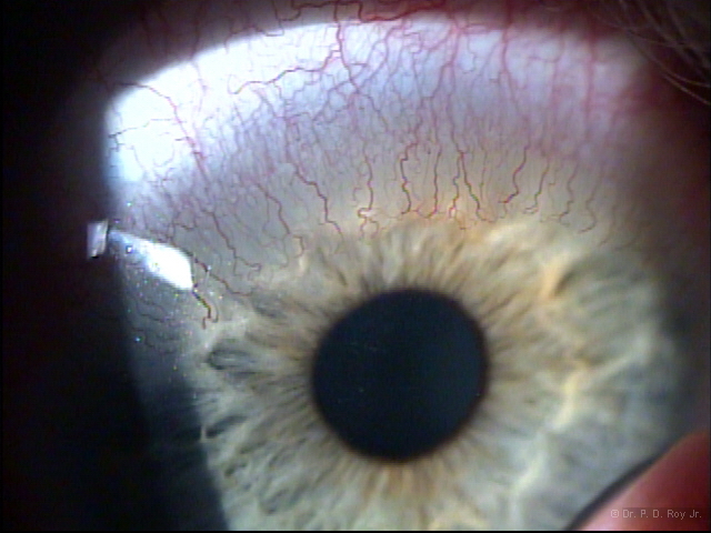
Neovascularization
Posterior Capsular Opacification
(after cataract)
This clouding forms behind the Intra-Ocular Lens (IOL).
.jpg)
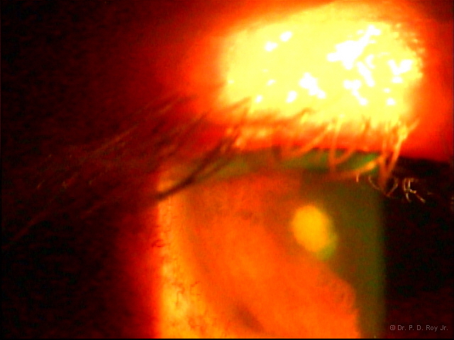
Ulcer
Pterygium
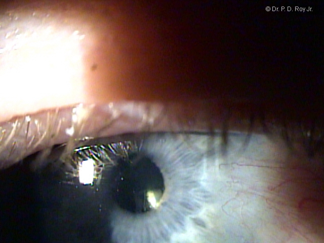
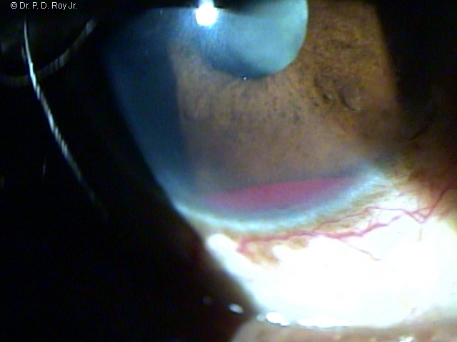
Blood in front chamber
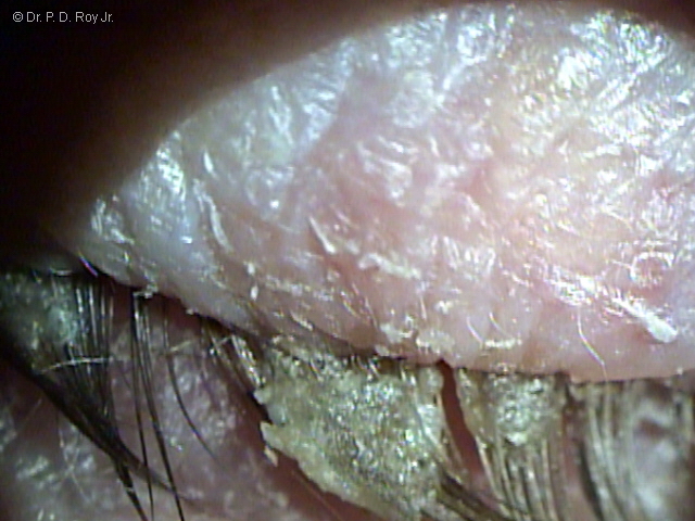
Blepharitis
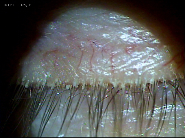
Blepharitis
Why REGULAR eye care and follow up is important.
Five photographs that show a growing nevus (freckle) that can turn into a Melanoma (cancer).
A growing nevus (freckle) can turn into a Melanoma (cancer). Small dark spot in sequence below as it grows larger.
2005
A growing nevus (freckle) can turn into a Melanoma (cancer). Small dark spot in sequence below as it grows larger.
2008
A growing nevus (freckle) can turn into a Melanoma (cancer). Small dark spot in sequence below as it grows larger.
2010
A growing nevus (freckle) can turn into a Melanoma (cancer). Small dark spot in sequence below as it grows larger.
2013
A growing nevus (freckle) can turn into a Melanoma (cancer). Small dark spot in sequence below as it grows larger.
2017
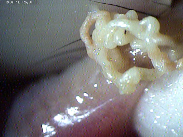
Oil Gland being expressed
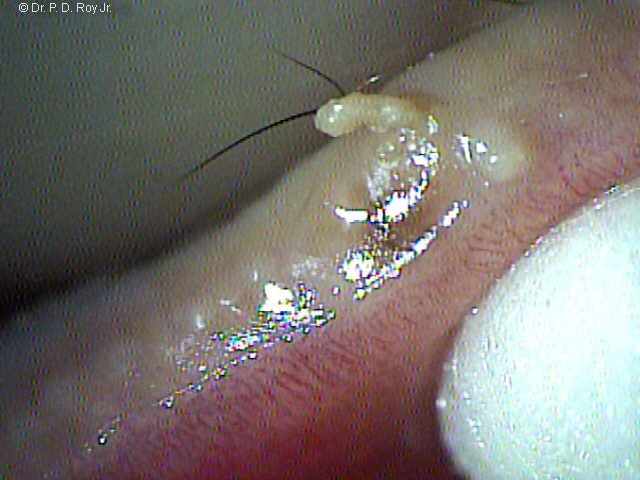
Oil Gland being expressed
Detached Retina
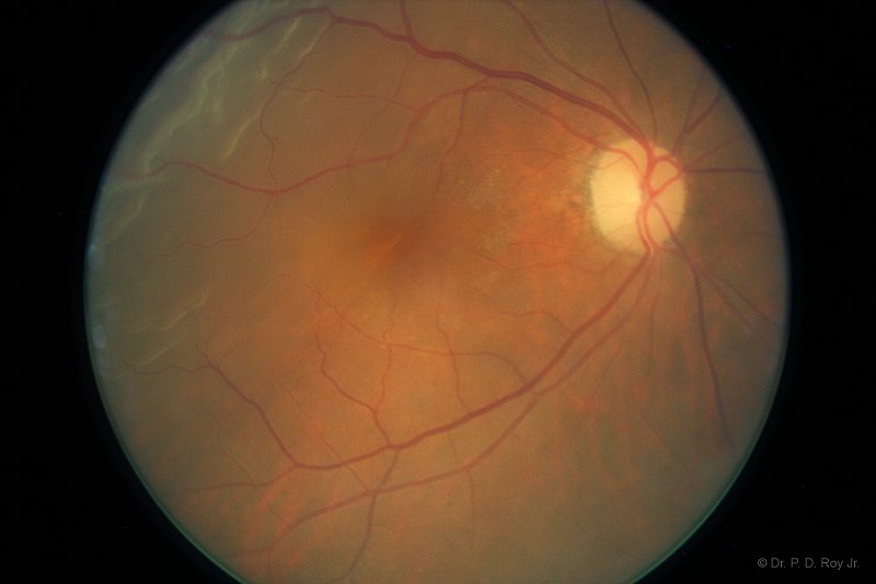
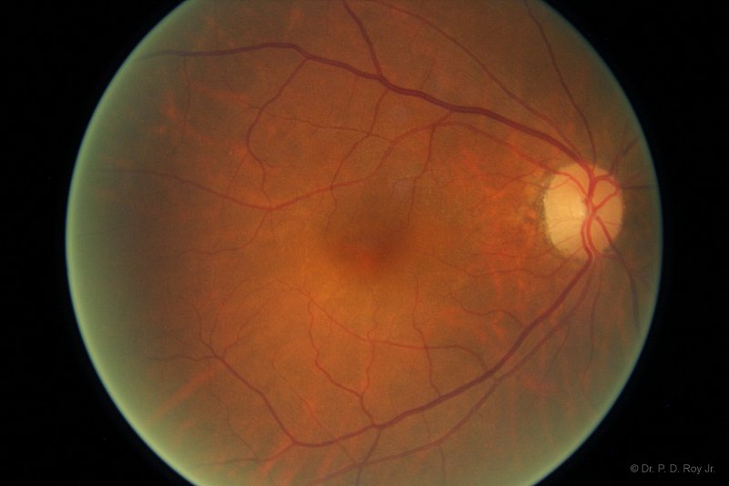
Detached Retina
After detachment repair five weeks out.
Photography by Dr. P. D. Roy, Jr. © All Rights Reserved.
Adenoviral Conjunctivitis
(known as shipyard fever in World War II)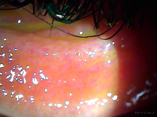
Adenoviral Conjunctivitis
(known as shipyard fever in World War II)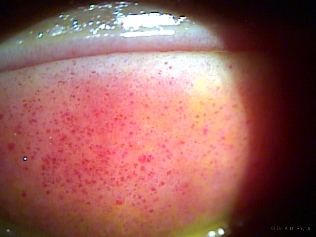
Bacteria Corneal Ulcer
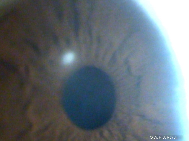
Bacterial Conjunctivitis
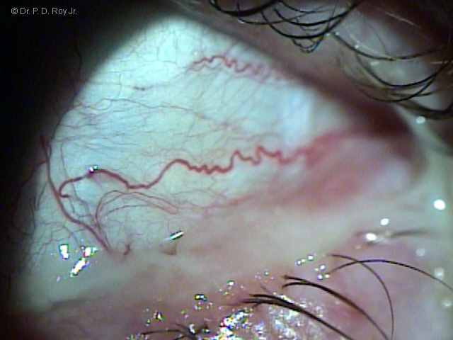
Photography by Dr. P. D. Roy, Jr. © All Rights Reserved.
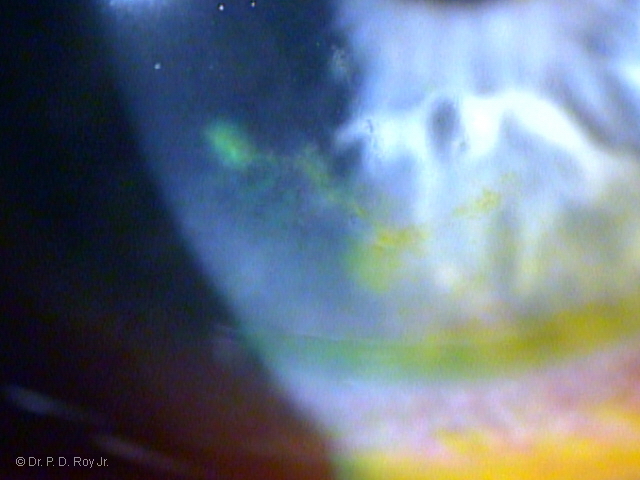
Viral Infection-Fever Blister
First Day Patient Presented
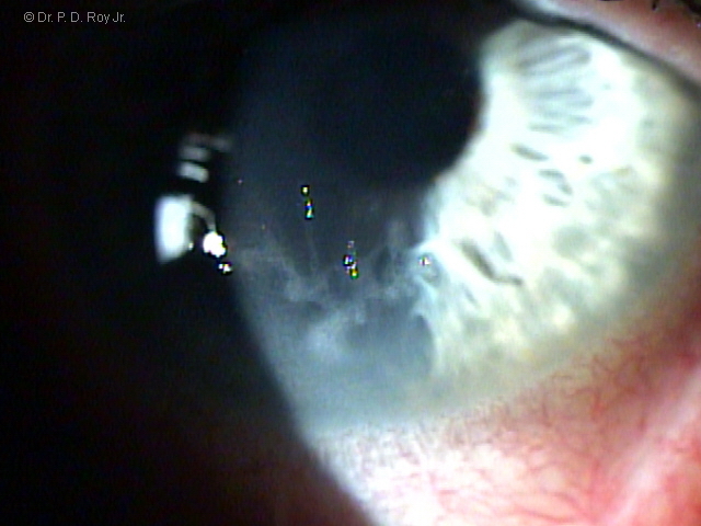
Viral Infection-Fever Blister
Third Day Patient Presented
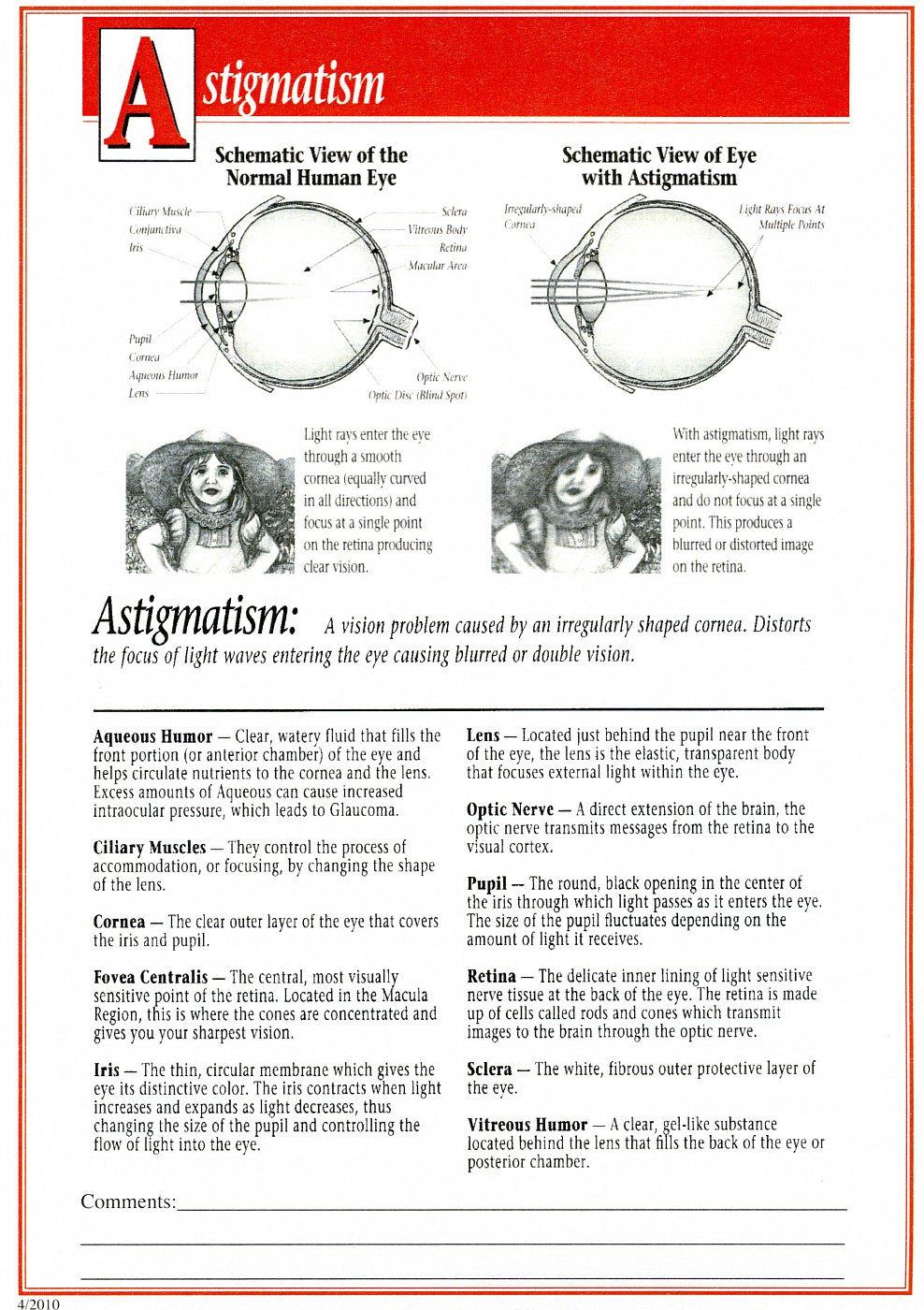
Additional Common Eye Disease Information
TESTIMONIAL
"Dr. Roy is the most thorough eye doctor that I have ever used. I now live in a neighboring state and drive 2 1/2 hours instead of trying to find someone here. He is very knowledgeable and has helped me to get through several scary things with my eyes. I recommend him to the highest degree. His office is small and his staff is friendly, courteous, helpful and professional. You won't be sorry that you use him for annual or ongoing optometric assistance." ~ Anonymous Patient, 5-Star Review, Ratemds.com
